Cultural heritage under the microscope
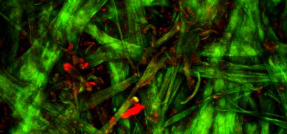
What do corneas, bones and skin have in common? The answer: collagen, a protein well known to researchers at the Optics and Biosciences Laboratory* (LOB) for its optical properties. This biological fiber is present in large quantities in the extracellular environment, providing the framework for living tissue. However, Gaël Latour, a teacher-researcher at the University of Paris-Saclay, who carries out his research at the LOB at the École Polytechnique, observes it outside the usual framework of biology. The physicist uses the laboratory's multi-photon microscope to assist heritage conservation-restoration professionals. “We're interested, for example, in parchments, which are nothing more than highly stretched animal skins, stripped of their fat and hair. This gives us a large collagen matrix... and a lot of work to do,” laughs the researcher.
Indeed, multi-photon microscopy, in which the LOB teams are specialists, is particularly sensitive to molecular organizations lacking a center of symmetry. And collagen, with its triple helices assembled into fibrils, which in turn are compacted into fibers, is an ideal candidate. On the parchments, the scientists observed signals identical to those seen on a cornea or the dermis of the skin - second harmonic generation (SHG) signals. To obtain them, the LOB's multi-photon microscope sends very brief infrared laser pulses onto the sample to be analyzed, and focuses its light beam on a very precise point. Thanks to the physical properties of collagen, a small fraction of this infrared light is reflected in the visible range. “If we observe SHG signals (in green on the images collected), it means that the collagen in our parchment is structured and in good condition. Degraded, it turns into gelatin, loses its hierarchical structure and sends back fluorescence signals (in red on the image)”, explains Gaël Latour.
An objective ally for heritage restorers
From now on, the researcher's job is to establish a scale of collagen degradation - and therefore of the parchment's state of conservation - based on images obtained using multi-photon microscopy. “To do this, we place contemporary parchments in an ageing chamber and observe them under the microscope at different stages. At the same time, we carry out conventional destructive analyses (editor's note: micro-samples subjected to calorimetric analysis and transformed into gelatin) with a metric that we know. Then we compare”. In this way, heritage restorers have at their disposal a fine, non-destructive analysis technique for the objects they treat, and even a mapping of their structure. It complements their expertise and enables them to adopt the right restoration protocols. This work is a godsend, since it provides an opportunity to better understand the optical signals obtained with collagen using multi-photon microscopy, and to feed fundamental research on the subject. “So much so, in fact, that a current thesis in the field considers parchment to be a model material for light-matter interactions”, stresses the researcher.
On the strength of this proof of feasibility, the technique has been adopted by the Centre de Recherche sur la Conservation at the Muséum national d'Histoire naturelle (MNHN) in Paris, which has acquired its own multi-photon microscopy platform as part of its work on analyzing and diagnosing the state of conservation of leather and parchment. “We're now turning our attention to cellulose, another non-centrosymmetric structure whose SHG signals it would be interesting to study during degradation”, says Gaël Latour. The scientist has already approached the Institut photonique d'analyse non-destructive européen des matériaux anciens (Ipanema) at the Soleil synchrotron. Traces of crystalline cellulose dating back 5,000 years have been discovered there on archaeological artefacts...
About:
Gaël Latour is a physicist specializing in optics, and a lecturer at the UFR Sciences of the Université Paris-Saclay (Orsay). He teaches physics, particularly optics, from L1 to M2. His research activities at the Laboratoire d'Optique et Biosciences de l'École polytechnique focus on the development of new imaging modalities, in particular non-linear optical microscopy, and their applications in the biomedical field (cornea, skin) and for the study of heritage objects
*LOB: a joint research unit CNRS, Inserm, École Polytechnique, Institut Polytechnique de Paris, 91120 Palaiseau, France


![[REFLEXIONS] Justice is key to successful energy transition](/sites/default/files/styles/actualite_liste/public/actualites/images/2026-01/Article%20_Energy%20Justice_%20-%20C%C3%A9line%20Guivarch.png?h=802ccbad&itok=atCAvrcB)
![[REFLEXIONS] The intertwined technical and political challenges of energy security](/sites/default/files/styles/actualite_liste/public/actualites/images/2026-01/Article%20Energy%20Security%20-%20Mathieu%20X%C3%A9mard.png?h=802ccbad&itok=BIpi1GC0)
![[Video] A look back at IP Paris Research Day 2025](/sites/default/files/styles/actualite_liste/public/actualites/images/2026-01/LOOK-BACK-2300x1075.png?h=aec66867&itok=vmLt9cUV)
![[REFLEXIONS] Breaking down the barriers to energy sustainability](/sites/default/files/styles/actualite_liste/public/actualites/images/2026-01/Article%20_Energy%20Sustainability_%20-%20Philippe%20Drobinski.png?h=802ccbad&itok=H6cZMnr7)
